Yesterday, I went to see a specialist called a neuro-opthamologist. We only have one in the Pacific Northwest, and it turns out that practices in Spokane, Wa (about 80 miles from where I live). She grew up in a little tiny town in eastern WA, Famington, and went to the University of Idaho as an undergrad, so I guess she decided to stay closer to home, and I am very grateful for that!
Since I had surgery, I have had a piece of my brain puzzle that was not clear, which has had to do with my vision. I’ve known that I could not see in a specific area (low and to the right), but I also have known that there was something about seeing certain things that was challenging . . . I have described it by saying that I have to “think to see” sometimes.
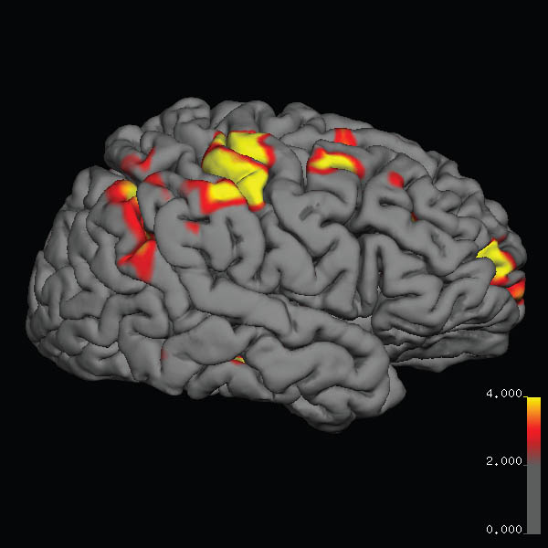
The testing that I had done yesterday explained what is going on. I have, as my doctor describes it, a “double-whammy” to deal with. I have damage to the part of my brain that sees the lower right side of the visual field in both eyes. But I also have damage to a part of the brain that tracks fast movements, like a baseball, or a cat flying by (not that my cats fly by quickly these days!). This area of the brain that tracks fast movement is in the left temporal lobe, and it would have been in the path of the surgical point of entry.
She explained to me that there isn’t a rehab program for restoring this part of the brain. But then, there wasn’t for the other parts of my brain that where compromised by many seizures, an A.V.M., and the surgery. So, my new challenge is how to work on rehabbing this. My choice of work will be a Nia technique called “head and eye” movement. It has to do with moving the arms around the body and tracking them with the eyes. This will help with the visual field limitation, as I need to learn to turn my head and look, in order to see what is low and to my right. I need to transform this action of turning my head to see, rather than attempting to see low and to the right with my eyes, which cannot do this. So with head and eye movements, I will practice turning my head in order to see.
I am also hoping that it will help with the tracking, as I vary the speed of tracking slow moving to quickly moving arms, hands, and fingers. While I’m not ready to have people throw baseballs at me to practice quick tracking, perhaps hanging out with a bunch of playful, quick-moving kittens? Or perhaps, just dancing with cats, as the image suggests?
I’ll keep you poster, and here’s a great piece on this part of the brain that I never knew about before yesterday. It’s amazing how specific certain brain functions can be! So send me good energy as I attempt to rehab my visual fast track center! In gratitude and joy for life force energy to continue healing, Kristine
http://www.livescience.com/29417-how-brain-tracks-moving-objects.html